
-
Package Including
- 1 * Handset
- 1 * 30xIris Lens
- 1 * Protective Lens Cover
- 1 * USB Cable
- 1 * IridologyChart
- 1 * User Manual
- 1 * Warranty card
- 1 * CD
- 1 * Aluminum Box
Iridology Camera Chart 12 MP HD 30X 9811U Iriscope Iris CCD Analysis Softw 3.31, NO Basement!
This is a professional 12MP family iriscope (eye exam camera with iris). As it is manual focus, its brightness and light focus can be adjusted by a wheel on the handle. It has a high resolution image sensor as we already mentioned and it is 12 megapixels, it has 5 quick access buttons and LED lights so that the exams are easy to perform and effective. It has a special DSP image sensor with a built-in optical image stabilizer.
12MP Iridology Camera Software and Operating System Compatibility
Compatible with Windows 7, 8, 10 and 11 and since 2021 we have a compatible version for MAC (Apple), I mean an independent Windows software, which works with MAC and a special usb or dongle for MAC. This iridology camera 12 MP (digital iriscope) includes a latest iris analysis software that is version 3.31 and checks the body conditions as well as help in the diagnosis and prevention of many ailments, in addition to analyzing the Sclera as we already mentioned.
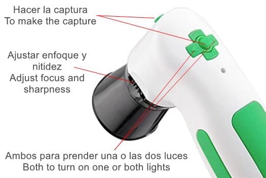
This 12MP iridology camera has a system of 5 buttons on top of its body and another on the side of the head, a total of 6 buttons of which only 4 work, since the two vertical ones (up and down) are replaced by the wheel that It is the one who exercises the function of adjusting the sharpness in the photo taking. The previous image explains how these 6 buttons work.
Other features of the 12MP Iridology Camera
- Automatic white balance and contrast adjustment.
- Dual image comparison function.
- 3D-Negative capture mode.
- It has a DSP image processor.
- Compatible with iris lenses, hair lenses and skin lenses.
- Maximum resolution: 2560 × 1920.
- Certification: FCC, CE.
- It is NOT compatible with IOS or MAC system.
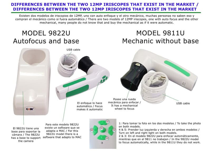
About the 12MP Iridology Camera with manual focus and no stand base.
This is a professional 12MP family iriscope (eye exam camera with iris). As it is manual focus, its brightness and light focus can be adjusted by a wheel on the handle. It has a high resolution image sensor as we already mentioned and it is 12 megapixels, it has 5 quick access buttons and LED lights so that the exams are easy to perform and effective. It has a special DSP image sensor with a built-in optical image stabilizer.
12MP Iridology Camera Software and Operating System Compatibility
Compatible with Windows 7, 8 and 10 and unfortunately it is not compatible with IOS MAC, although its similar 12MP model 9822U is compatible with MAC . This 12MP iridology camera (digital iriscope) includes the latest iris analysis software, and checks body conditions as well as aids in the diagnosis and prevention of many ailments.
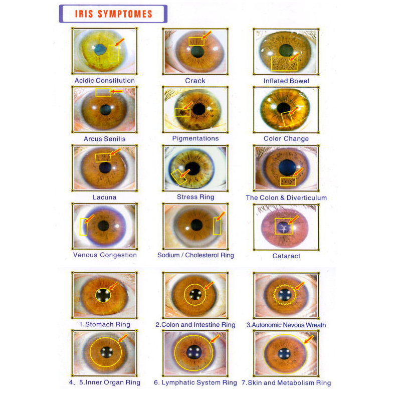
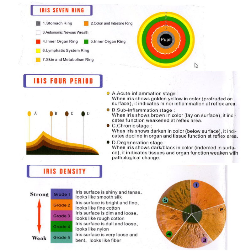
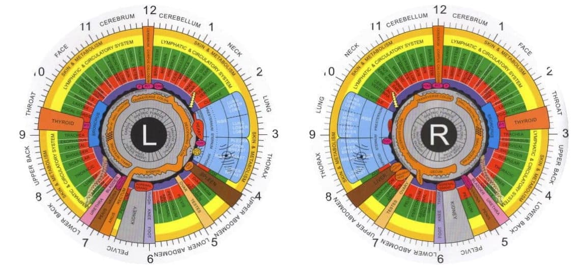
Everything that you could evaluate or analyze with this iriscope idology 12MP at 30X
|
|
|
|
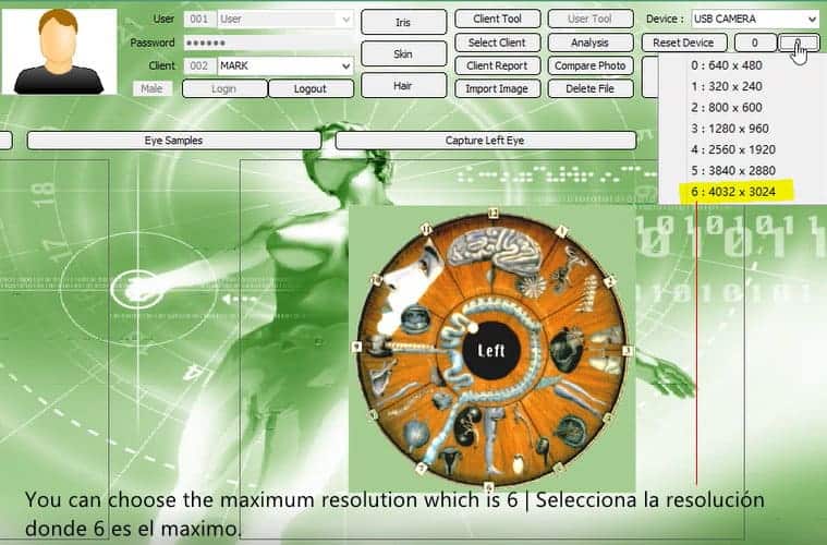
For best results with the iriscope iridology 5MP you should set the camera to the maximum resolution which is 4032 x 3024 which corresponds to grade 6.
After placing the camera at 4032 x 3024, which corresponds to grade 6, we will begin with the capture of the iris and once we have them, just select the eye we want and proceed first with analysis and then with the car that is the one we are going to choose.
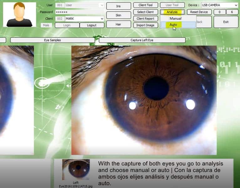
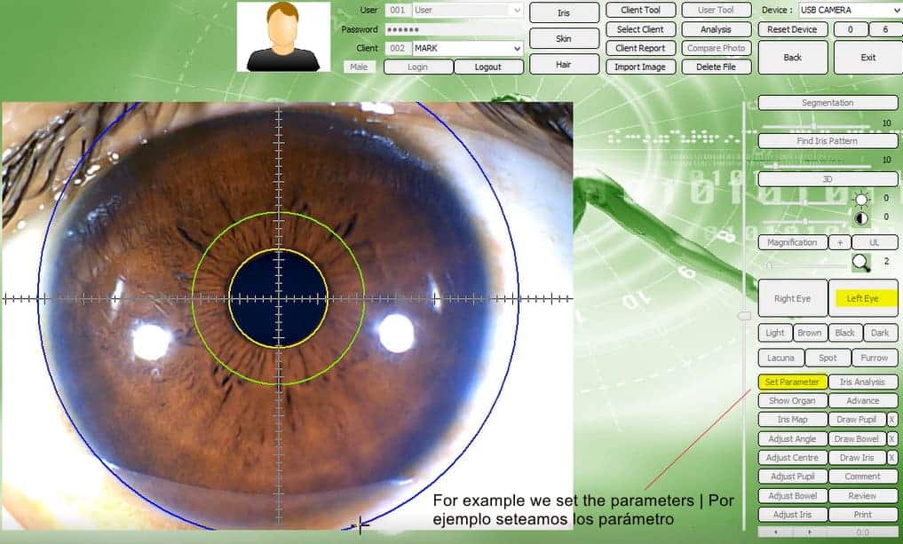
Once you select the eye to analyze, we must set the parameter and begin to mark it throughout the area of the eye including the pupil with which we started first. In the video it is explained better.
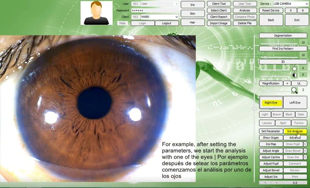
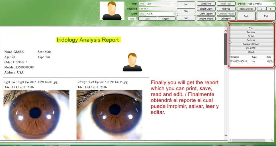
After taking the captures of both eyes, you only have to select the one you want to analyze and click on analyze iris, it is important that you check the box show organ.
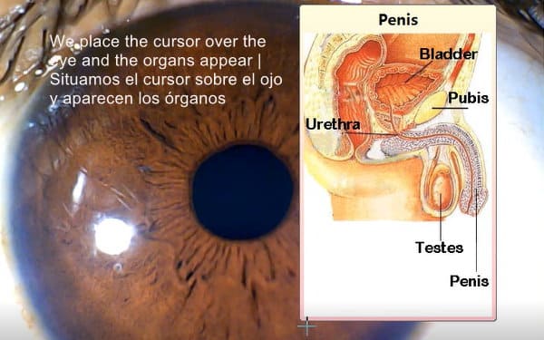
When you check the box show organs they will appear in your analysis as you can see in the following image.
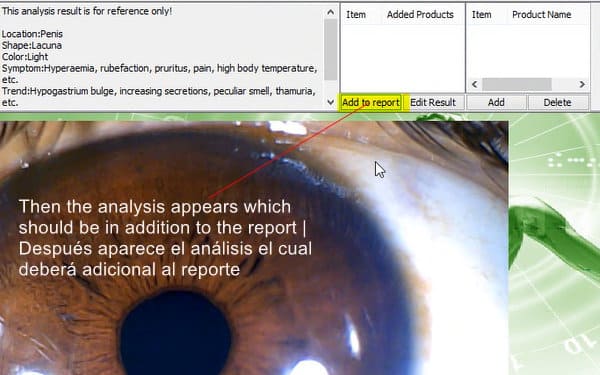
Once the organ appears and the program will analyze that area and a report will appear in a box which I show below, you just have to add it to the report and that's all.
The reports will appear as you can see them in the following image, you can print, save and do with them what the menu as shown allows you.
This is just an idea of what you can do with our 12MP USB iriscope or iridology camera, it is obvious that to use it you know something about iridology, if you do not have knowledge, we recommend that you take a course that there are many options on the internet, and so you can get the most out of our iriscope.
For your peace of mind, the breakage margin of an iriscope of ours is 0.001% and its functionality and veracity is 99% , I tell you this so that you do not look for objections or excuses because there are none, more than 1000 units have been sold and the returns they do not reach 10 units, and all returns have simply been, because the person who bought them did not know even 1% of iridology.
* The products and information found on this website are not to replace any medical or health professional advice as the information displayed is for informational purposes only. Results may vary. These statements have not been evaluated by the Food and Drug Administration (FDA). Therefore our supplements and equipment are not intended to diagnose, prevent, treat or cure any disease or health problem. Always check with your doctor or healthcare professional before proceeding with any new diet, exercise regimen, supplement use, or if you have any concerns about a health problem. We are not responsible for grammatical errors that the page shows. Product availability, prices and promotions may be changed without prior notice.